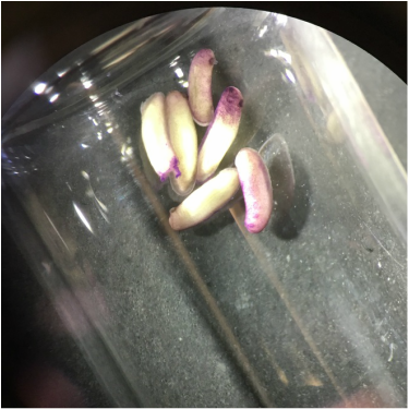
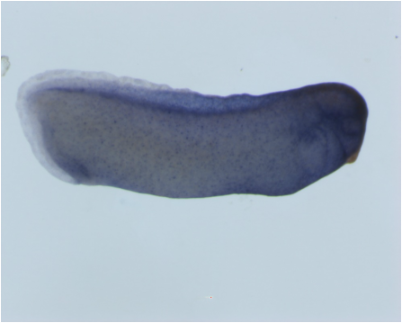
This image was one I found to be very cool as I was actually able to see some staining in some of the later-stage embryos after B-gal injection. You can see the colored areas in the embryos meaning that the injections were successful as we have also bee working on microinjection of embryos with Beta-galactosidase as well as GFP. I took this image while looking through the microscope to check on the staining of our embryos, and it was very fulfilling to see that it worked!
Since working in the lab, I have learned many different techniques regarding imaging of the embryos. We use Xenopus embryos, and one important aspect of the work we do is being able to take images that depict the expression of what we are interested in. In one of my first few weeks working in the lab, I learned how to use the Zeiss V12 Discovery stereoscope to study and take photographs of the embryos either in bright field or under fluorescence. In Figure 1 above, you can see one of the images that I took using this stereoscope. I have also learned how to process these images using the ZEN imaging software, which is a very cool program that assists in getting great images of the embryos!
|
Author: Megen WittlingI am a second year Biology Major. I started working in the lab in January. ArchivesCategories |