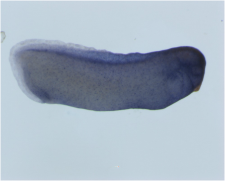
Figure 1: Image of Embryo I Took Using Stereoscope and ZEN Imaging Software
Since working in the lab, I have learned many different techniques regarding imaging of the embryos. We use Xenopus embryos, and one important aspect of the work we do is being able to take images that depict the expression of what we are interested in. In one of my first few weeks working in the lab, I learned how to use the Zeiss V12 Discovery stereoscope to study and take photographs of the embryos either in bright field or under fluorescence. In Figure 1 above, you can see one of the images that I took using this stereoscope. I have also learned how to process these images using the ZEN imaging software, which is a very cool program that assists in getting great images of the embryos!
 RSS Feed
