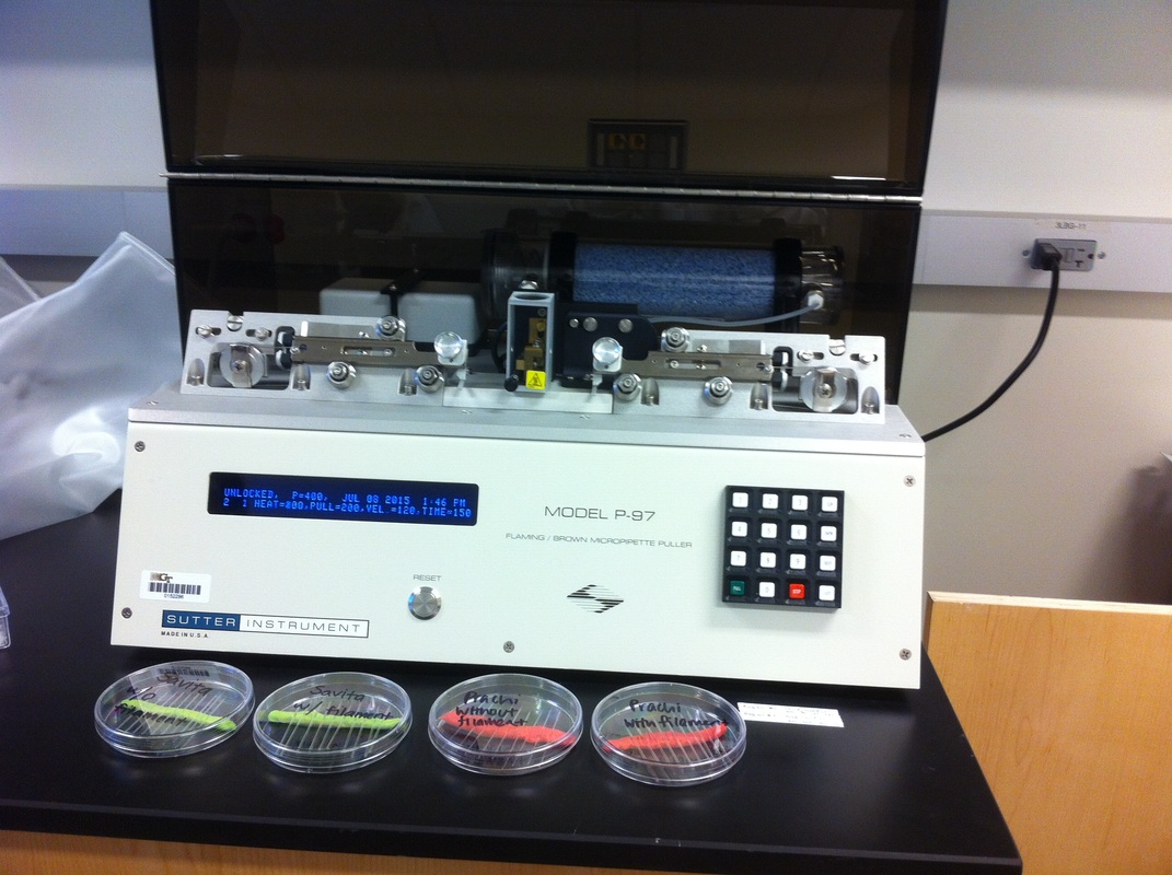
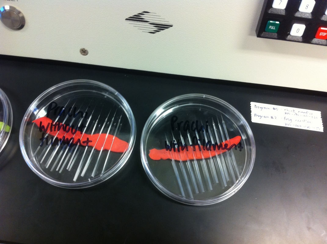
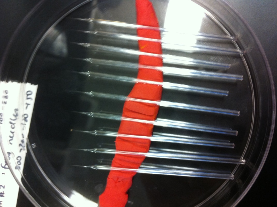
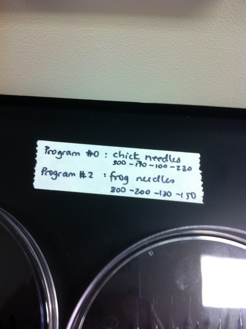
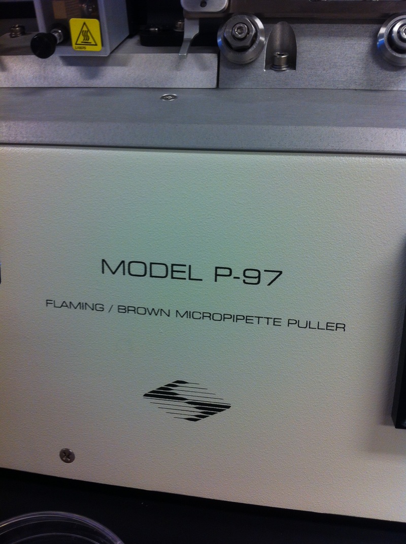
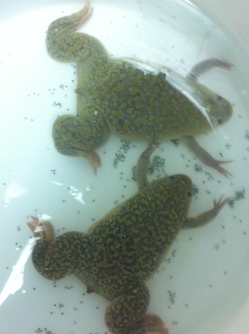
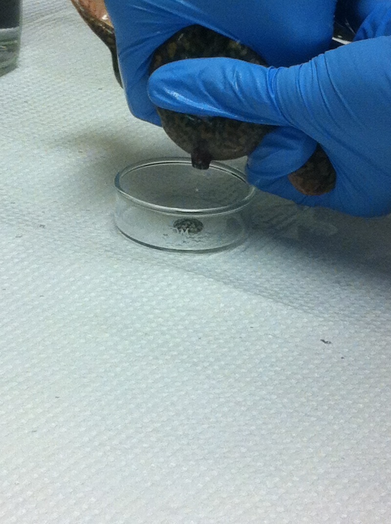
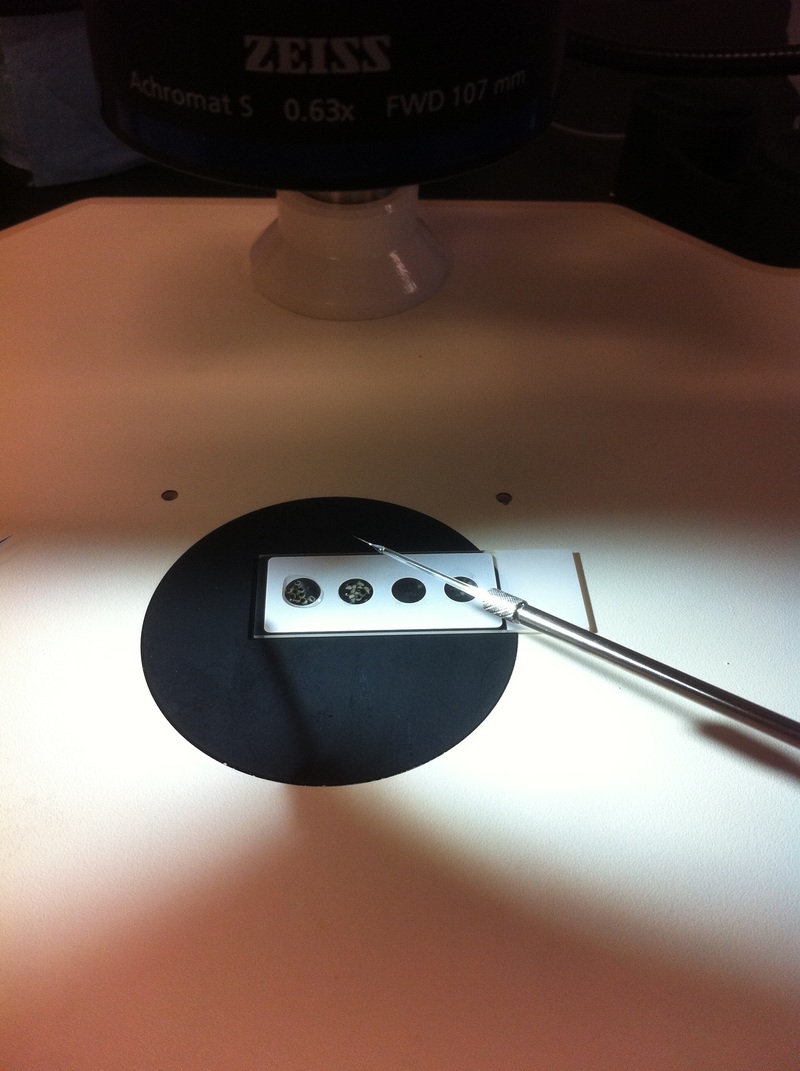
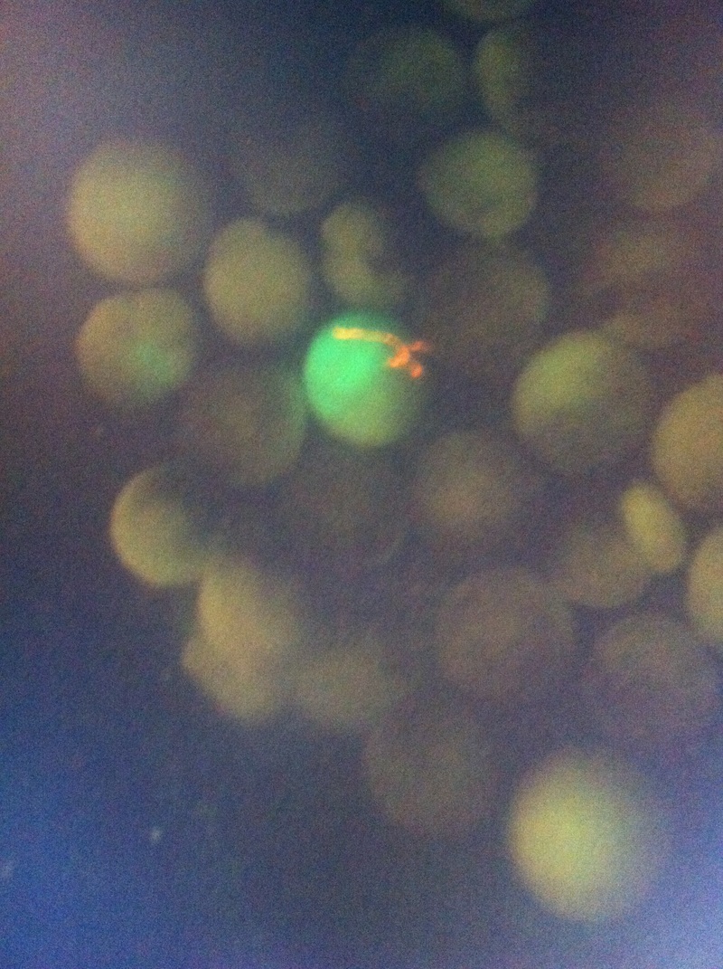
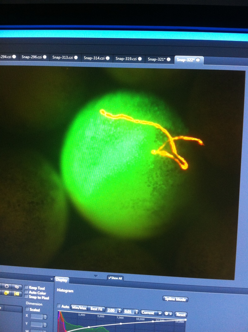
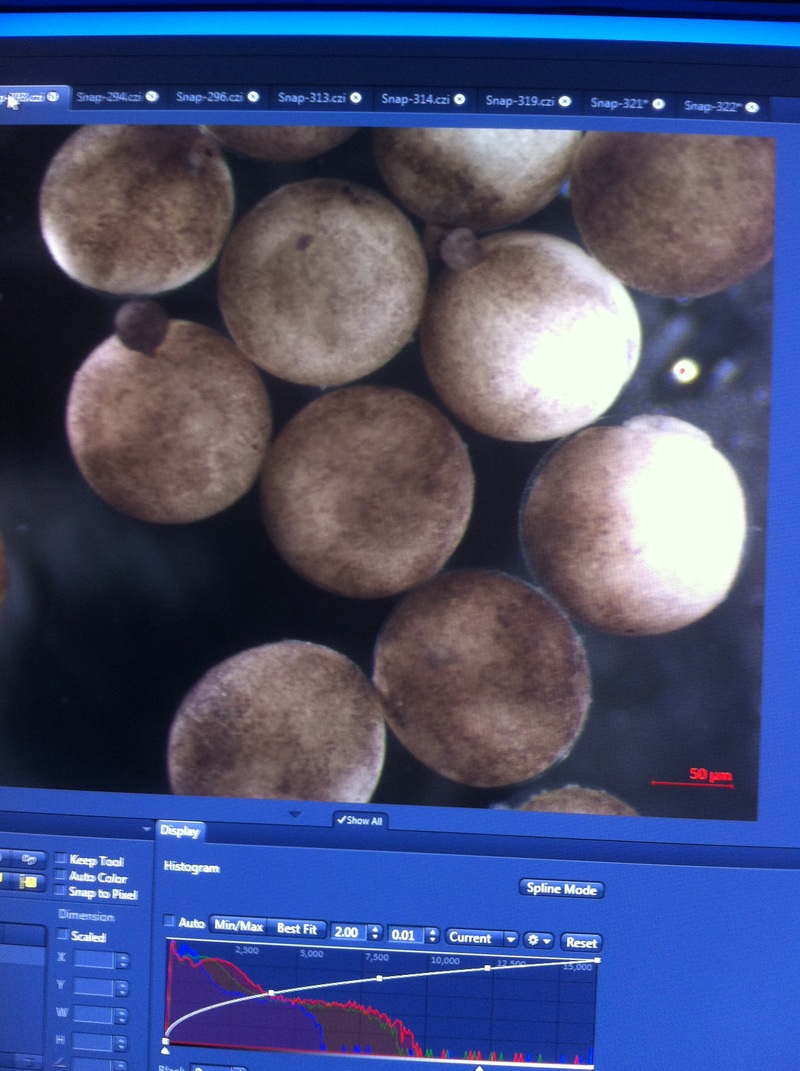
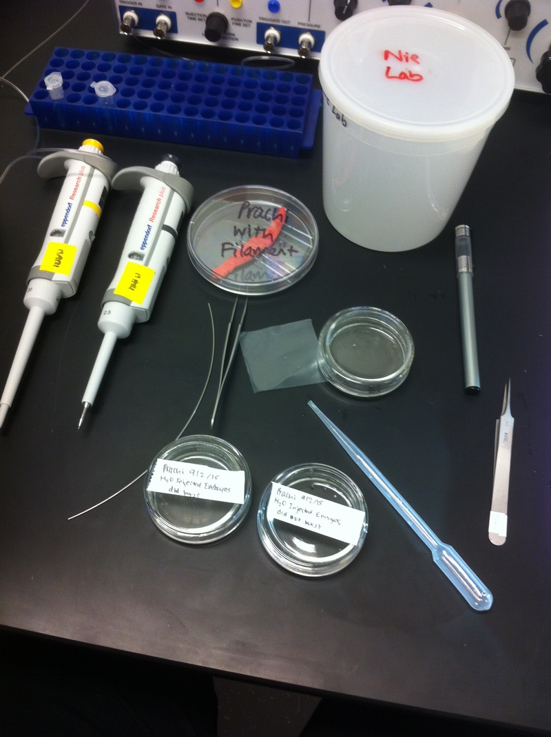
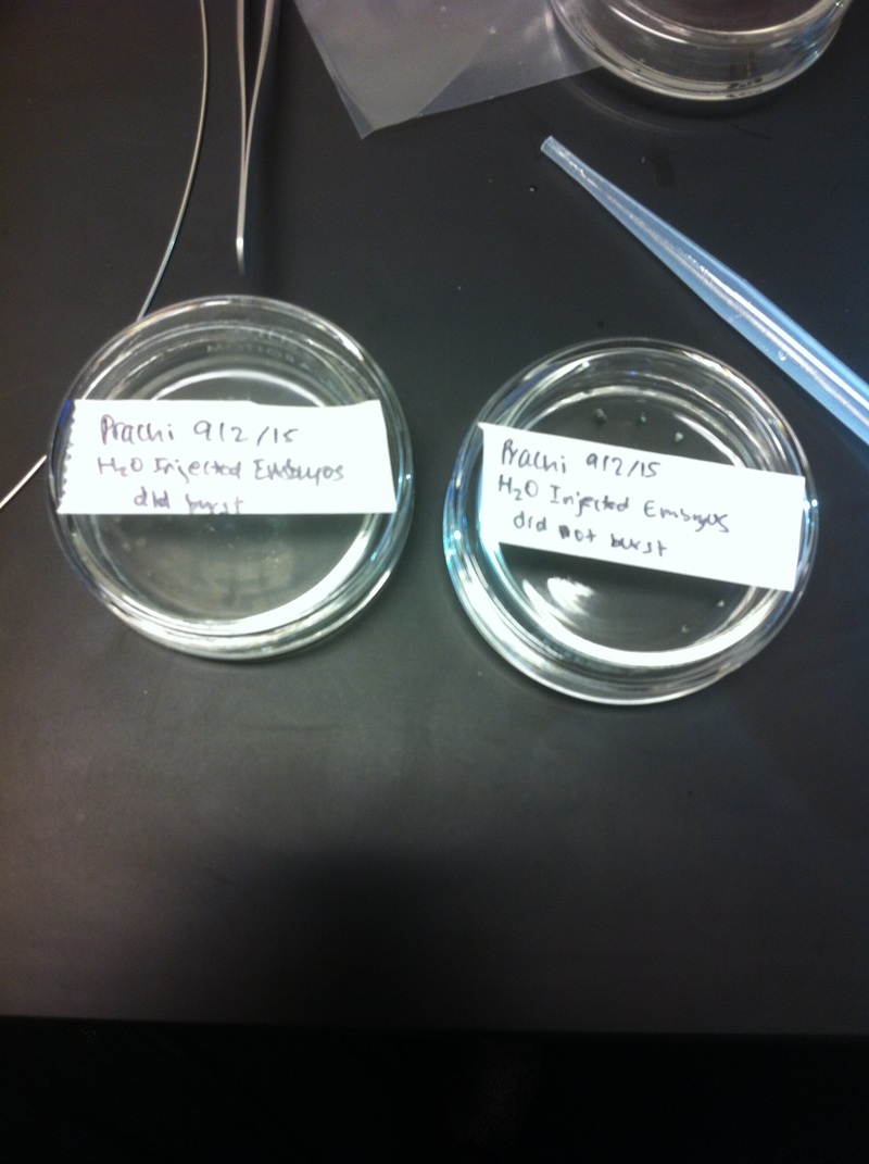
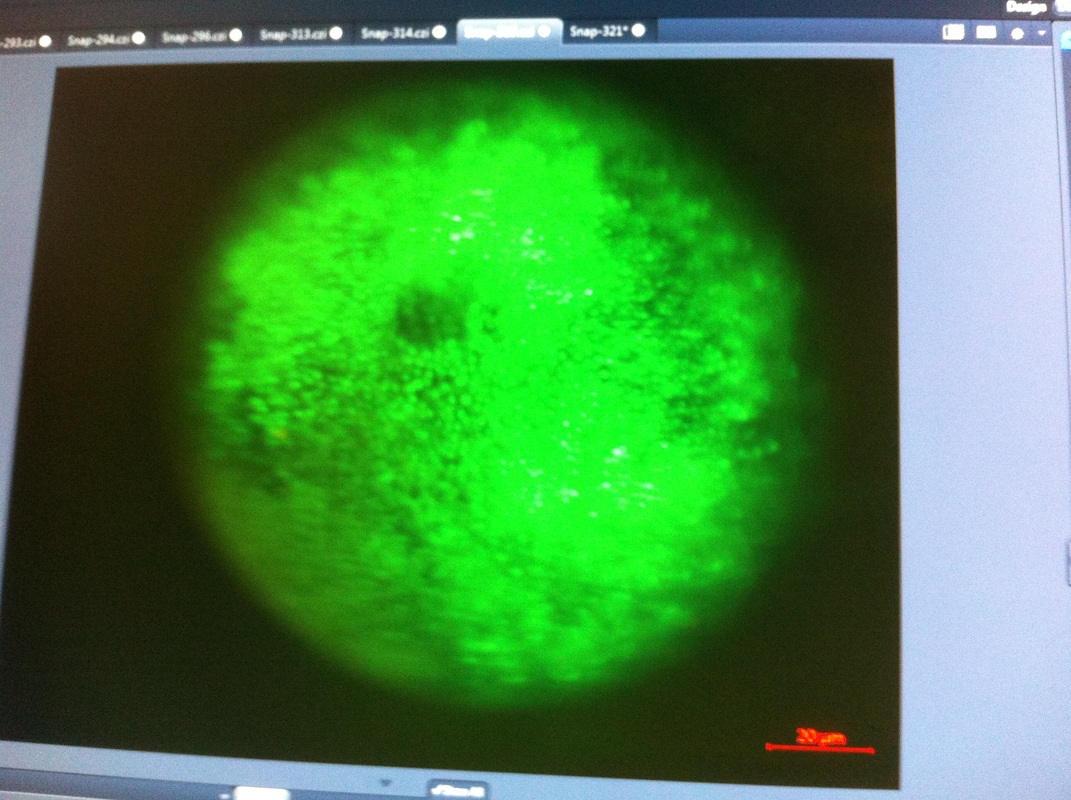
Being a part of Nie Lab has definitely been an awarding experience. After spending a month in the lab, I have learned about multiple protocols and various techniques on how to successfully carry out each task. The first major protocol that I learned was RNA injection in Xenopus laevis embryos. As an undergraduate researcher, I was astounded that I would be able to inject frog embryos. Previous laboratories never allowed me to be involved in something so delicate and precise. Before I started injecting RNA into the embryos, Mariana did a few demonstrations to make sure that I understood the details and the larger purpose of this procedure: to see if certain genes are expressed during the embryos' early developmental stages. The entire process takes a lot of preparation beforehand. Mariana had to prime the frogs with hormones the night before to ensure that there would be eggs in the morning, which were later fertilized with male testis, and finally stored in a petri dish with 3% Ficoll solution after they had been placed in 3% Cysteine solution to dejelly. Before I began the procedure for the first time, I extracted approximately twenty needles from the Model P-97 Micropipette Puller. I chose program #2 which was designated for frog needles. Needle extraction took about ten to fifteen minutes in duration. Afterwards, I firmly slid the needle in place of where the metal rod had been, cut the very tip of the needle with forceps, and began to calibrate the machine with water. After the calibration, I collected the RNA with my needle and began to inject the frog embryos slowly and delicately. It was extremely difficult to match what I was doing with my needles while I was squinting into the microscope. My hand-eye coordination was not too skillful, and several of the embryos burst and made the slides cloudy. Many times, my needle would break and I'd have to immediately replace it with a new needle. Although frustrating at first, I became better at injecting after multiple days in the lab with lots of practice. Afterwards, we took pictures of the Xenopus laevis embryos under UV light to detect if the RNA had successfully been injected. My success rate was not too high, but one fluorescent embryo sparked hope and determination for the next time I would inject embryos. Attached are some pictures from my experience with RNA injection.
-Prachi Mishra

 RSS Feed
