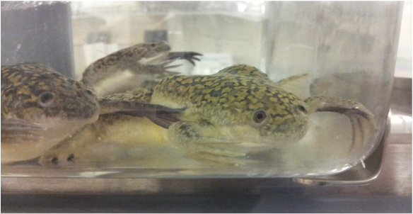
Xenopus laevis (African Clawed Frog)
Frogs of the genus Xenopus are one of the two model organisms we use in the lab to study neural crest cell migration. All species of Xenopus are completely aquatic, meaning they can spend their entire lives in the water and never have to come up on land. Unlike the frogs we commonly think of, Xenopus do not have tongues to catch insects. Instead, they used their front paws to move food into their mouths. In the wild, they are scavengers and will eat almost anything organic that they find in their ponds. Like many species of fish, these frogs have lateral lines, which consists of clusters of cells along the sides of the animal that can detect vibrations and help them find food in murky water.
The species of Xenopus we use in the lab is Xenopus laevis, commonly known as the African Clawed Frog. This species is often used in developmental biology research due to its large embryos that are easy to work with. In the Nie lab, we are interested in the movement of neural crest cells during the development of X. laevis embryos. In order to track the cells, we focus on genes that are expressed during their migration and visualize their gene products using in situ hybridization, a technique that makes proteins visible under ultraviolet light.
The species of Xenopus we use in the lab is Xenopus laevis, commonly known as the African Clawed Frog. This species is often used in developmental biology research due to its large embryos that are easy to work with. In the Nie lab, we are interested in the movement of neural crest cells during the development of X. laevis embryos. In order to track the cells, we focus on genes that are expressed during their migration and visualize their gene products using in situ hybridization, a technique that makes proteins visible under ultraviolet light.
Gallus gallus domesticus (Domesticated Chicken)
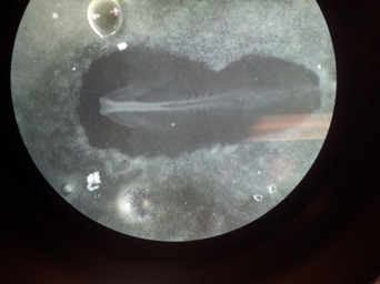
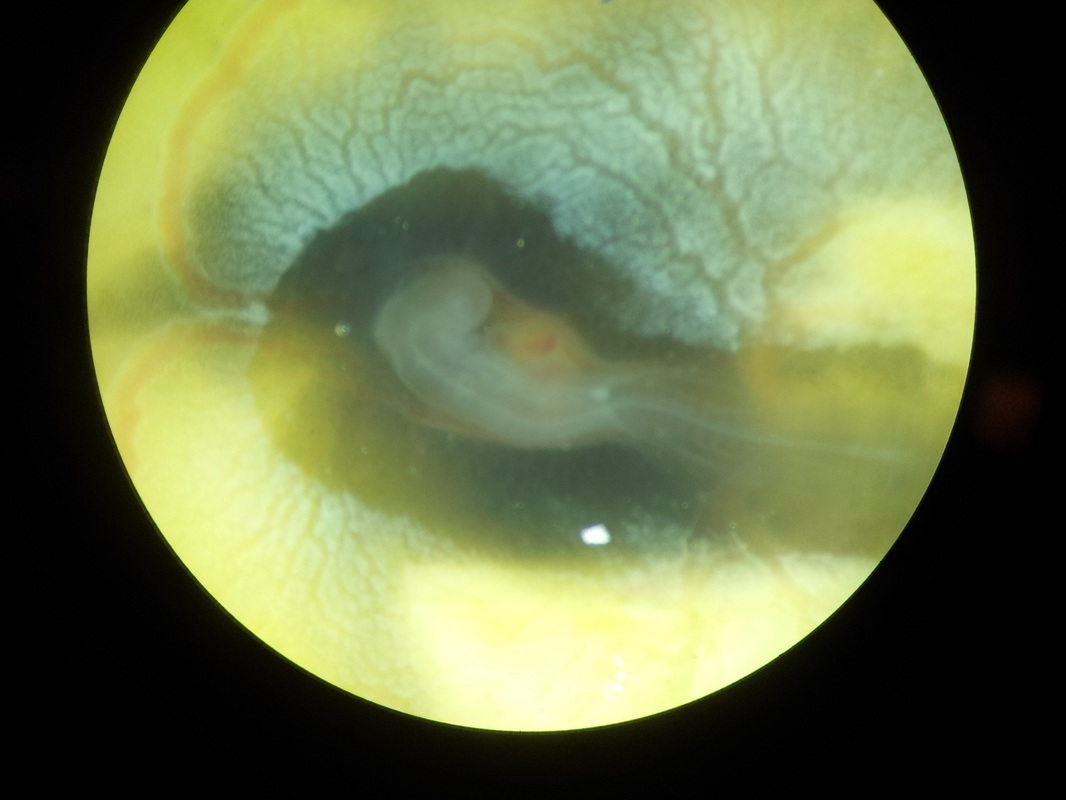
The second model organism we use in the lab is the chicken, or more precisely fertilized chick embryos in their eggs. By cutting open the egg shell, it is possible to remove the embryos from the yolk without damaging them. It is then possible to conduct in situ hybridization or immunostaining to detect the gene products that are expressed by neural crest cells during their migration. The images below show chick embryos as seen through a dissecting microscope. The one on the right is in a more advanced state of development and has a visible heart beat (the heart is the red spot to the right of the head, above the abdomen).
 |  |
-Tobias Hoffmann
 RSS Feed
