An in situ hybridization was performed in order to gauge the effects of EDTA treatment on the migration of neural crest cells, as seen through staining for the expression of the standard neural crest marker, sox10. A control group was established, along with an experimental group in which embryos were treated with 1mM EDTA at three different stages of development. After treatment, embryos were fixed around stage 30. With EDTA acting as a chelating agent that would degrade the function of the MMP-14 protein product through removal of the zinc ion of its active site, this in situ would help to isolate MMP-14’s influence over neural crest cells. The probe for sox10, a gene ubiquitous to NC cells, was used to determine the proper migration of these cells and the development of structures arising from it, and thus used to infer the impact of ineffective MMP-14 protein operation. This in situ hybridization was conducted from June 20th to June 24th.
Hypothesis:
Embryos treated with 1mM EDTA will feature a decreased expression of sox10 within neural crest- derived structures upon being stained, indicating a decreased migration and subsequent specification of neural crest cells due to MMP-14 inhibition by EDTA.
Results:
Hypothesis:
Embryos treated with 1mM EDTA will feature a decreased expression of sox10 within neural crest- derived structures upon being stained, indicating a decreased migration and subsequent specification of neural crest cells due to MMP-14 inhibition by EDTA.
Results:
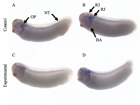
Figure 1. In situ hybridization with sox10 antisense probe. A: Stage 30. B: Stage 27. C: Stage 30. D: Stage 30. All views are lateral with dorsal at the top and the anterior to the left. Experimental embryos C and D were treated with 1mM EDTA at the stages 8, 14, and 22 (not differentiated in image). OP, otic placode; NT, neural tube; R3, rhombomere 3; R5, rhobmomere 5; BA, branchial arches.
Discussion:
Most distinguishable in this staining is the otic placode, the blue ring near the anterior-dorsal region of the embryo; this will become the future ear of the developed frog. Neural crest cells that migrate to this area and contribute to the development of the inner ear are cranially-derived. No apparent difference in expression can be seen between the control and experimental embryos in regards to sox10 expression in the otic placode. The most significant difference in expression may be seen in the streams corresponding to the 3rd and 5th rhobomere of the hindbrain. Within the experimental embryos, these streams have a decreased expression and did not migrate as successfully down the dorsal-ventral axis of the embryo in comparison to the controls. The branchial arches are also lacking expression within the experimental embryos. Expression along the dorsal neural tube appears to be unaffected.
Conclusions:
These experimental results support the hypothesis that MMP-14 protein inhibition through EDTA treatment negatively impacts the expression of the neural crest marker sox10, shedding light on its role in the proper migration of neural crest cells. In future experiments, it would be beneficial to fix the embryos at an earlier stage (stage 24-26), as neural crest activity and the expression of gene associated with them begins to decline and decrease in area of expression at later stages (such as around stage 30). With EDTA being a fairly blunt and unspecific method of countering the activity of the MMP-14 protein, future experiments will be using a specific chemical inhibitor to influence MMP-14 and its functions on, just as EDTA, a protein-level basis.

 RSS Feed
