EDTA is a multidentate ligand that may act as a chelating agent in the removal of zinc ions from the active site of the MMP-14 protein product. With MMP-14 being largely dependent on the presence of this metal ion in order to digest collagen of the extracellular matrix, the addition of EDTA would very likely inhibit the proper migration and subsequent specification of cells reliant on MMP-14, namely neural crest cells. I performed a simple experiment in which different stages of embryos were treated with different concentrations of EDTA in order to observe any macroscopic effects on development before an in-situ hybridization is conducted using a standard neural crest marker. This experiment was conducted from June 7th to June 9th.

EDTA is a hexadentate ligand, and thus creates six bonds with the zinc ion within MMP-14's binding site in order to remove it and render the protein inactive.
Hypothesis:
With greater concentration of EDTA in which the embryos develop, greater physical malformations and a greater death rate will be observed. EDTA treatment will have the greatest effect on the youngest embryos that it is added to, as embryos are most sensitive to environmental conditions during early development and these will have the longest span of time to be exposed before being fixed.
Experimental Design:
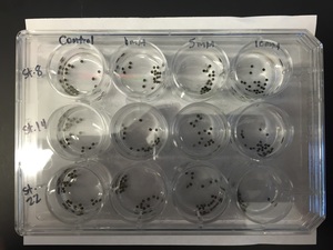
A 12-well plate was used to contain the embryos. Three stages were tested: stages 8, 14, and 22. Results from stage 8 would demonstrate the effect of EDTA on neural crest cells when it is added prior to specification, stage 14 would demonstrate the effect of EDTA on specified but pre-migratory neural crest cells, and stage 22 would demonstrate whether EDTA was toxic to embryos in general. Four concentrations of EDTA were applied to these three stages: 0 mM (control), 1mM, 5mM, and 10mM. These concentrations were diluted in 0.1x MMR. In each well, embryos were kept in 3mL of solution and 20 embryos were originally placed within each of the 12 wells, with dead embryos being removed as they appeared.
With greater concentration of EDTA in which the embryos develop, greater physical malformations and a greater death rate will be observed. EDTA treatment will have the greatest effect on the youngest embryos that it is added to, as embryos are most sensitive to environmental conditions during early development and these will have the longest span of time to be exposed before being fixed.
Experimental Design:
A 12-well plate was used to contain the embryos. Three stages were tested: stages 8, 14, and 22. Results from stage 8 would demonstrate the effect of EDTA on neural crest cells when it is added prior to specification, stage 14 would demonstrate the effect of EDTA on specified but pre-migratory neural crest cells, and stage 22 would demonstrate whether EDTA was toxic to embryos in general. Four concentrations of EDTA were applied to these three stages: 0 mM (control), 1mM, 5mM, and 10mM. These concentrations were diluted in 0.1x MMR. In each well, embryos were kept in 3mL of solution and 20 embryos were originally placed within each of the 12 wells, with dead embryos being removed as they appeared.
Day 1 (6/7/16):
Embryos were collected and fertilized. Experimental set-up was established and, upon reaching stage 8, EDTA treatment was applied to the first row of embryos. Samples were stored at 15°C overnight.
Day 2 (6/8/16):
Initial observations of treated stage 8 embryos: embryos within the control and 1mM wells appeared to be gastrulating properly and were healthy. Embryos within the 5mM and 10mM were not gastrulating properly and eventually died off, with the pigment of the animal pole appearing as scattered black pinpoints and the vegetal pole being a cloudy white. This result was expected, as embryos are the most sensitive during gastrulation, and thus it was likely that the two higher concentrations of EDTA would result in embryo death.
The embryos of the second and third rows were gastrulating normally.
Upon reaching stage 14, EDTA treatment was applied to the second row. Within 30 minutes of application, approximately one-fourth of the embryos within the 5mM and 10mM wells were starting to die off, appearing a cloudy white. The embryos treated with 1mM EDTA, however, appeared healthy. Samples were stored at 15°C overnight.
Day 3 (6/9/16):
Observations of treated stage 8 embryos: controls appeared to be healthy with no abnormalities. Embryos treated with 1mM appeared less healthy, having some surface damage and with uneven pigment. These embryos, compared to their controls, had a stunted development and had not elongated properly. All embryos of the 5mM and 10mM wells had died, having a dark animal pole and cloudy white vegetal pole.
Observations of treated stage 14 embryos: controls appeared to be healthy with no abnormalities. Embryos treated with 1mM EDTA had a stunted development and had not elongated properly, similar to the same treatment of the stage 8 embryos. All of the embryos treated with 5mM and 10mM had died off, having a cloudy grey color.
Upon reaching stage 22, EDTA treatment was applied to the last row of embryos. Within 20 minutes of treatment, a distinct pattern was observed in about half of embryos within the 10mM well. These embryos demonstrated cell death beginning at their posterior end. The cells would turn a cloudy white, the posterior end of the embryo would then burst, the embryo would contort within its vitelline membrane, and the embryo would then die. This eventually took place in almost all embryos within this treatment, leaving only four living by stage 24. Interestingly, this trend did not take place in the embryos treated with 5mM; they appeared healthy compared to the controls. Thus, the controls, 1mM, and 5mM embryos appeared healthy upon reaching stage 24.
The embryos were then fixed and dehydrated in preparation for an in-situ hybridization using a probe for sox10.
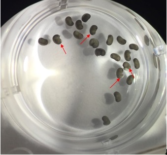
The interesting pattern of cell death observed in the stage 22 embryos treated with 10mM EDTA.
Conclusions:
While more tests need to be conducted, it does appear that there exists a negative relationship between EDTA concentrations and embryo survivorship and morphological normalcy. While I initially believed that 5mM and 10mM might have been too large of a concentration to get beneficial results without killing the embryos, the fact that some embryos treated at stage 22 survived both of the higher concentrations might indicate that the role of metalloproteinases (which are inhibited by the EDTA) may be more vital in the early development of the embryo, such as during gastrulation and neurulation. However, if this experiment were to be repeated, I would extend the time in which the stage 22 embryos were exposed to the EDTA, as these embryos could have likely died as well had they not been fixed only a couple stages after their treatment.
Images:
http://openwetware.org/wiki/EDTA
 RSS Feed
